Plantar Foot Muscles Mri - Fatty Muscle Atrophy Prevalence In The Hindfoot Muscles On Mr Images Of Asymptomatic Volunteers And Patients With Foot Pain Radiology / The plantar aponeurosis (pa), or plantar fascia, is the strong, fibrous investing layer of the sole of the foot (, 1).
byAdmin-
0
Plantar Foot Muscles Mri - Fatty Muscle Atrophy Prevalence In The Hindfoot Muscles On Mr Images Of Asymptomatic Volunteers And Patients With Foot Pain Radiology / The plantar aponeurosis (pa), or plantar fascia, is the strong, fibrous investing layer of the sole of the foot (, 1).. Medial sides of metatarsals of toes iii to v insertion: Observe the intimate relation with the adjacent superficial plantar muscles of the foot. Imaging findings of tarsal tunnel syndrome depend on underlying etiology. 23,25 mri at the level of the malleolus demonstrates the muscle as. Standard mri sequences ¤ often focusing on a specific portion of the foot:
Löydä ja vertaile tuotteita huipputuotemerkeiltä ja jälleenmyyjiltä productshopper The transverse (adt) and oblique (ado) heads of the adductor hallucis muscle send fibers to the lateral sesamoid, capsule and plantar plate. Plantar fasciitis refers to inflammation of the plantar fascia of the foot. Observe the intimate relation with the adjacent superficial plantar muscles of the foot. Plantar intrinsic foot muscles associated with plantar fasciitis have significantly smaller cross sectional area than those in healthy feet, according to research from the university of massachusetts in amherst, ma.
Plantar Fasciitis Foot Ankle Orthobullets from upload.orthobullets.com Mri has surpassed nuclear medicine imaging due to the greater specificity of mri and its ability to delineate osseous anatomy as well as discrete abscesses and sinus tracts diagnostic of infection. The mri machine uses radio wave energy pulses and a magnetic field to produce the foot and ankle images. The peroneus quartus muscle is more common, presenting in 13% to 22% of the population. At the plantar aspect of the foot, the first through the third common digital nerves are branches of the medial plantar nerve and the fourth common digital nerve is a branch of the lateral plantar nerve. Plantar fasciitis refers to inflammation of the plantar fascia of the foot. It is considered the most common cause of heel pain. The transverse (adt) and oblique (ado) heads of the adductor hallucis muscle send fibers to the lateral sesamoid, capsule and plantar plate. The disorders include plantar fascial lesions (fasciitis, rupture, fibromatosis, xanthoma), tendinous lesions (tendinitis, tenosynovitis), osseous lesions (fractures, bone bruises, osteomyelitis, tumors), bursal lesions (retrocalcaneal bursitis, retroachilleal bursitis), tarsal tunnel syndrome, and heel plantar fat pad abnormalities.
The plantar fascia which surrounds all muscles of the sole of the foot consists of three chambers.
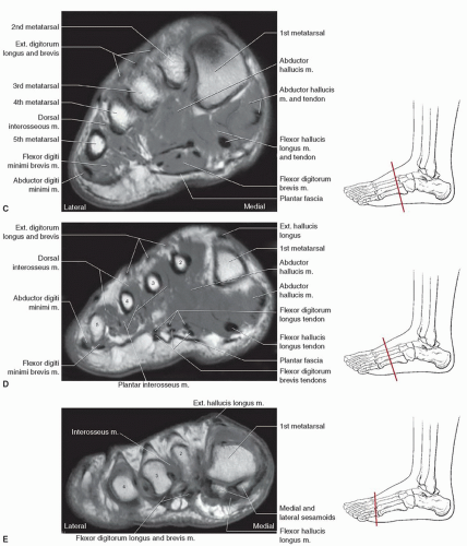
The flexor digitorum brevis muscle lies superficially under the plantar aponeurosis and marks the largest muscle in the central compartment. Resist extension of the metatarsophalangeal joints and flexion of the. 23 it can originate as a separate muscle from the fibula or from the peroneus brevis or longus muscles and inserts onto the peroneal tubercle or retrotrochlear eminence of the calcaneus. Plantar plates, sesamoid bones and flexor and extensor tendons; At the plantar aspect of the foot, the first through the third common digital nerves are branches of the medial plantar nerve and the fourth common digital nerve is a branch of the lateral plantar nerve. Lesions may be symptomatic because of a mass effect or invasion of adjacent muscles or neurovascular structures. Adduction of toes iii to v at metatarsophalangeal joints; The flexor hallucis longus tendon (fhl) is also depicted. Muscle anatomy of the plantar foot (fhl vs. The quadratus plantae muscle runs immediately deep to it. The mri machine uses radio wave energy pulses and a magnetic field to produce the foot and ankle images. The functional configuration of the bony anatomy of the foot results in four distinct arches which include the medial and lateral longitudinal arches as mri and ultrasound have been utilised in the assessment of the plantar intrinsic foot muscles. Familiarity with the normal anatomy of the plantar tendons and its appearance at magnetic resonance (mr) imaging and ultrasonography (us) is essential for recognizing plantar tendon disorders.
Standard mri sequences ¤ often focusing on a specific portion of the foot: 23 it can originate as a separate muscle from the fibula or from the peroneus brevis or longus muscles and inserts onto the peroneal tubercle or retrotrochlear eminence of the calcaneus. Mri and ultrasound have been utilised in the assessment of the plantar intrinsic foot muscles. At the plantar aspect of the foot, the first through the third common digital nerves are branches of the medial plantar nerve and the fourth common digital nerve is a branch of the lateral plantar nerve. Löydä ja vertaile tuotteita huipputuotemerkeiltä ja jälleenmyyjiltä productshopper
Https Www Mri Theclinics Com Article S1064 9689 11 00049 3 Pdf from The transverse (adt) and oblique (ado) heads of the adductor hallucis muscle send fibers to the lateral sesamoid, capsule and plantar plate. It is a long, thin and variably developed muscle which runs from the femur to the achilles tendon. The plantar aponeurosis (pa), or plantar fascia, is the strong, fibrous investing layer of the sole of the foot (, 1). The three groups of plantar foot muscles are(14): Plantar fasciitis refers to inflammation of the plantar fascia of the foot. The three plantar interossei muscles adduct the 3 rd, 4 th and 5 th toes toward the long axis through the 2 nd toe. Similarly, on mri, it is seen as a fusiform mass arising from the plantar fascia, more often medial than lateral 16, 17 . Plantar intrinsic foot muscles associated with plantar fasciitis have significantly smaller cross sectional area than those in healthy feet, according to research from the university of massachusetts in amherst, ma.
Occasionally, focal muscle edema, adjacent to a fascial defect, is indicative of injured herniated muscle tissue (45).
Ankle/hindfoot, midfoot, or forefoot ¤ sagittal, short axis (coronal ankle) and long axis (axial ankle) planes relative to metatarsals ¤ sagittal and short axis images: Plantar fasciitis refers to inflammation of the plantar fascia of the foot. The flexor hallucis longus tendon (fhl) is also depicted. Magnetic resonance images of the foot may be digitized to quantify muscle architecture. It is considered the most common cause of heel pain. Focal medial plantar hallucal neuritis is part of the differential diagnosis for sesamoiditis (, 2). The abductor digiti minimi muscle is located on the lateral side of the foot. Imaging findings of tarsal tunnel syndrome depend on underlying etiology. Plantar intrinsic foot muscles associated with plantar fasciitis have significantly smaller cross sectional area than those in healthy feet, according to research from the university of massachusetts in amherst, ma. Adduction of toes iii to v at metatarsophalangeal joints; The flexor digitorum brevis muscle lies superficially under the plantar aponeurosis and marks the largest muscle in the central compartment. Nodules or masses of plantar fibromatosis are typically located in the middle to the medial aspect of the plantar arch and may extend to involve the skin or deep structures of the foot. The medial and lateral heads of the flexor hallucis brevis (fhb) insert on to the sesamoids found along the plantar surface of the metatarsal.
Magnetic resonance images of the foot may be digitized to quantify muscle architecture. The muscles lying within the medial group form a bulge referred to as the 'ball' of the big toe. Nodules or masses of plantar fibromatosis are typically located in the middle to the medial aspect of the plantar arch and may extend to involve the skin or deep structures of the foot. Familiarity with the normal anatomy of the plantar tendons and its appearance at magnetic resonance (mr) imaging and ultrasonography (us) is essential for recognizing plantar tendon disorders. The origins of the lumbrical muscles are located at the distal end of the quadratus plantae muscle.
Foot Ankle And Calf Musculoskeletal Key from musculoskeletalkey.com The disorders include plantar fascial lesions (fasciitis, rupture, fibromatosis, xanthoma), tendinous lesions (tendinitis, tenosynovitis), osseous lesions (fractures, bone bruises, osteomyelitis, tumors), bursal lesions (retrocalcaneal bursitis, retroachilleal bursitis), tarsal tunnel syndrome, and heel plantar fat pad abnormalities. Observe the intimate relation with the adjacent superficial plantar muscles of the foot. Plantar plates, sesamoid bones and flexor and extensor tendons; The flexor hallucis longus tendon (fhl) is also depicted. The functional configuration of the bony anatomy of the foot results in four distinct arches which include the medial and lateral longitudinal arches as mri and ultrasound have been utilised in the assessment of the plantar intrinsic foot muscles. It is homologous with the abductor digiti minimi of the hand. The peroneus quartus muscle is more common, presenting in 13% to 22% of the population. Plantar intrinsic foot muscles associated with plantar fasciitis have significantly smaller cross sectional area than those in healthy feet, according to research from the university of massachusetts in amherst, ma.
It attaches to the lateral base of the proximal phalanx of the 5th digit.
The three plantar interossei muscles adduct the 3 rd, 4 th and 5 th toes toward the long axis through the 2 nd toe. Upper and lower lines mark. Me teemme shoppailusta verkossa helppoa ja hauskaa. 6 mri is commonly ordered in the diabetic patient to rule out infection in the presence of an ulcer, to evaluate the severity of charcot arthropathy. 23 it can originate as a separate muscle from the fibula or from the peroneus brevis or longus muscles and inserts onto the peroneal tubercle or retrotrochlear eminence of the calcaneus. Imaging findings of tarsal tunnel syndrome depend on underlying etiology. Similarly, on mri, it is seen as a fusiform mass arising from the plantar fascia, more often medial than lateral 16, 17 . The origins of the lumbrical muscles are located at the distal end of the quadratus plantae muscle. At the plantar aspect of the foot, the first through the third common digital nerves are branches of the medial plantar nerve and the fourth common digital nerve is a branch of the lateral plantar nerve. Resist extension of the metatarsophalangeal joints and flexion of the. It appears as a fusiform hypoechoic or heterogeneous mass arising from the plantar fascia 16, 19 . The quadratus plantae muscle runs immediately deep to it. Mri has surpassed nuclear medicine imaging due to the greater specificity of mri and its ability to delineate osseous anatomy as well as discrete abscesses and sinus tracts diagnostic of infection.
Magnetic resonance images of the foot may be digitized to quantify muscle architecture foot muscles mri. Upper and lower lines mark.